Scope of Practice

Surgical Expertise

Dr. Morgan manages a broad referral caseload across orthopedic, neurologic, soft tissue, thoracic, and minimally invasive surgery. Cases vary considerably in complexity; the following outlines his principal areas of practice.

Orthopedic Surgery

Bones, joints, tendons, and ligaments — from common referral conditions to complex reconstructive cases

Dr. Morgan manages orthopedic referrals ranging from cranial cruciate ligament disease and patellar luxation to complex periarticular fractures and salvage procedures. Preoperative CT and digital planning are used selectively when they are expected to improve surgical accuracy or change management.

TPLO — Tibial Plateau Leveling Osteotomy Cranial cruciate ligament disease, partial or complete tears; concurrent meniscal evaluation at arthroscopy or arthrotomy
Patellar Luxation Surgery Tibial tuberosity transposition, trochleoplasty, imbrication — graded approach based on severity
Fracture Repair Plating, interlocking nails, external skeletal fixation; diaphyseal, metaphyseal, and periarticular fractures
Elbow Disease Fragmented medial coronoid process (FMCP), osteochondrosis dissecans (OCD), un-united anconeal process; evaluated by CT and often managed with arthroscopy
Shoulder and Hip Disorders OCD of the shoulder, medial shoulder instability, femoral head and neck ostectomy (FHNO), total hip consultation
Tendon and Ligament Repair Achilles tendon injuries, biceps tenotomy/tenodesis, collateral ligament repair
Angular Limb Deformity Correction Corrective osteotomy for antebrachial and tibial deformities; preoperative CT planning
Joint and Bone Biopsy / Mass Excision Aggressive bone lesions, periosteal masses, joint pathology requiring histopathology
TPLO planning radiograph Femur fracture repair — before and after radiograph

Left: TPLO planning radiograph. Right: Before-and-after radiographs demonstrating internal fixation of a comminuted femoral shaft fracture.

Neurologic & Spinal Surgery

Surgical management of spinal cord compression and neurologic disease in dogs and cats

Neurologic surgery requires precise localization of the lesion, appropriate imaging, and careful surgical decompression. MRI is the imaging modality of choice for soft tissue and cord evaluation; CT myelography may be used in selected cases. Surgical timing and the degree of neurologic deficit are central to decision-making.

Hemilaminectomy Thoracolumbar IVDD (Hansen Type I and II); spinal cord decompression and disc material removal
Ventral Slot Procedure Cervical IVDD; ventral approach to the cervical disc space for decompression
Mini-Hemilaminectomy / Foraminotomy Selected thoracolumbar and lumbosacral lesions; foraminal decompression
Spinal Fracture / Luxation Stabilization Vertebral body plating, screw-rod constructs, pin-PMMA fixation for traumatic spinal instability
Lumbosacral Decompression Dorsal laminectomy and foraminotomy for cauda equina syndrome and degenerative lumbosacral stenosis
Selected Extradural Compressive Lesions Extradural masses with spinal cord compression amenable to surgical decompression; case-dependent approach
MRI spine multiview demonstrating IVDD at T13-L1

MRI multiview demonstrating thoracolumbar disc extrusion with spinal cord compression. This modality provides soft tissue resolution essential for surgical planning in neurologic cases.

All neurologic surgical cases benefit from thorough neurologic examination and lesion localization before imaging. The imaging choice — CT myelogram versus MRI — depends on clinical presentation, available anesthesia risk, and the suspected nature of the lesion.

Soft Tissue & Oncologic Surgery

Abdominal, gastrointestinal, hepatobiliary, urinary, and oncologic procedures

Soft tissue surgery encompasses a wide range of abdominal and body wall procedures. Oncologic surgery requires consideration of tumor margins, staging, and whether surgical excision improves prognosis or quality of life. Dr. Morgan performs both complex abdominal surgeries and targeted mass removal procedures in collaboration with oncology and internal medicine colleagues at VERG Brooklyn.

Gastrointestinal Surgery Intestinal resection and anastomosis, enterotomy, foreign body removal, gastric surgery, GDV/bloat
Splenectomy Splenic masses, splenic torsion, trauma; often urgent or emergency presentation
Hepatobiliary Surgery Liver lobectomy, hepatic mass removal, cholecystectomy, biliary surgery
Urinary Tract Surgery Cystotomy, urethrostomy, ureteral surgery, bladder mass resection
Oncologic Mass Excision Wide excision of soft tissue sarcomas, mast cell tumors, and other subcutaneous and intracavitary masses
Thyroid and Endocrine Surgery Thyroidectomy, adrenalectomy (open and laparoscopic), parathyroid surgery
Perineal and Reproductive Surgery Perineal hernia repair, perianal tumors, reproductive tract surgery
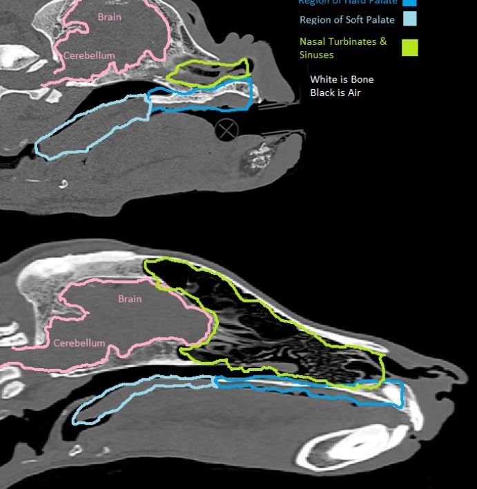
Nasal and Oral Surgery Nasal tumor debulking, mandibular and maxillary mass excision, nasal plane surgery
CT demonstrating splenic mass Annotated CT of nasal tumor

Left: CT demonstrating a splenic mass. Right: Annotated CT of a nasal tumor — useful for surgical planning and radiation therapy mapping.

Thoracic & Airway Surgery

Pulmonary, mediastinal, pericardial, pleural, and upper airway procedures

Thoracic surgery requires careful perioperative management and is typically performed with specialist anesthesia. Preoperative imaging — usually CT — is strongly encouraged for thoracic mass lesions to characterize anatomy and guide resectability before surgery. A dedicated page on chylothorax surgical management is available for referring veterinarians.

Lung Lobectomy Primary pulmonary tumors, lung lobe torsion, abscess or consolidation, thoracoscopic or open approach
Chylothorax Surgery Thoracic duct ligation, subphrenic pericardectomy, and cisterna chyli ablation — triple-approach management
Pericardectomy Subtotal or partial pericardectomy for pericardial effusion and constrictive pericarditis; thoracoscopic approach where appropriate
Laryngeal Paralysis (Tie-back) Unilateral arytenoid lateralization; typically indicated for moderate-to-severe upper airway obstruction
Brachycephalic Obstructive Airway Syndrome (BOAS) Nares widening, staphylectomy/palatoplasty, tonsillectomy, everted saccule removal; combined approach
Mediastinal Mass Excision Thymic masses and other anterior mediastinal lesions; approach determined by CT anatomy and resectability
Thoracic Trauma Management Pneumothorax, hemothorax, rib fractures, diaphragmatic hernia — stabilization and surgical repair
Tracheal Collapse — Surgery Consultation Extraluminal ring prosthetics for cervical tracheal collapse in selected patients
3D CT reconstruction of a thoracic mass

Color 3D CT reconstruction demonstrating the extent and vascular relationships of a thoracic mass. CT is strongly recommended before thoracic surgery to characterize the lesion and assess resectability.

Minimally Invasive Surgery

Arthroscopy, laparoscopy, and thoracoscopy

Minimally invasive techniques are used when they provide a meaningful clinical advantage for the individual patient — improved visualization, reduced morbidity, or faster recovery. They are not applied indiscriminately. Some conditions are better managed with open surgery, and the choice between techniques is part of the preoperative discussion.

Arthroscopy — Stifle Meniscal evaluation, OCD treatment, joint lavage; often combined with TPLO
Arthroscopy — Elbow FMCP treatment, OCD treatment, joint evaluation; can be combined with biceps tenotomy or tenodesis
Arthroscopy — Shoulder OCD treatment, medial shoulder instability, synovial biopsy
Laparoscopic Gastropexy Prophylactic stomach tacking (pexy) in high-risk breeds, typically performed at the time of spay or neuter; reduced morbidity versus open approach
Laparoscopic Liver Biopsy Hepatic sampling for hepatopathies; superior visualization compared with ultrasound-guided percutaneous biopsy
Laparoscopic Cryptorchidectomy Intra-abdominal retained testicle removal; avoids open celiotomy
Laparoscopic Adrenalectomy Adrenal masses; laparoscopic approach for selected cases based on size and anatomy
Thoracoscopy Lung lobectomy, pericardectomy, pericardioscopy, pleural biopsy; selected thoracic procedures
Arthroscopy in the operating room Laryngoscopy / scope image

Emergency & Trauma Surgery

Urgent surgical intervention for life-threatening injuries and conditions

VERG Brooklyn operates a 24-hour emergency and specialty hospital. Emergency surgical coverage involves the entire surgical and emergency team, not any single surgeon. Urgent surgical cases are assessed and prioritized based on clinical stability, injury severity, and the need for immediate intervention versus temporization.

For emergencies: Please call VERG Brooklyn directly at (718) 522-9400. The hospital operates around the clock. Emergency triage is coordinated by the emergency medicine and surgical teams.

Traumatic Fracture Repair High-energy fractures from motor vehicle accidents, falls, or penetrating trauma; stabilization and definitive repair
Penetrating Trauma Gunshot wounds, bite wounds, impalement injuries; exploration, debridement, and repair
Abdominal Emergencies Splenic hemoabdomen, gastrointestinal perforation, mesenteric volvulus, uroabdomen, septic abdomen
Gastric Dilatation-Volvulus (GDV) Decompression, gastric repositioning, and gastropexy; urgent intervention required
Gastrointestinal Obstruction Foreign body obstruction, intussusception — enterotomy or intestinal resection as indicated
Thoracic Emergencies Pneumothorax, hemothorax, diaphragmatic hernia, thoracic wound — stabilization and surgical management
Gunshot wound case — evaluation and repair

Penetrating trauma case. Emergency surgical evaluation, wound exploration, and repair. Images published without patient-identifying information.

Refer a Surgical Case

Contact VERG Brooklyn to discuss a referral or arrange imaging review.